loose body histology
This is the next level of organization in the human body up from cells themselves and a good. Backgroundhistologically based analyses of the nature and origin of loose bodies occurring in osteoarthrosis have been few and further study is warrantedmethodswe.

Xmlinkhub
J Orthop Sci 2006 11607613 DOI 101007s00776-006-1065-2 Original article Histologic classification of loose bodies in osteoarthrosis Koichi Saotome Kazuya Tamai.

. The small freely mobile loose bodies can be. Terminology Use of the term loose is frowned upon by some because the fragments do not. Definition general Defined as infestation caused by body louse Pediculus humanus corporis Skin lesions due to direct bite hypersensitivity reaction and itching related.
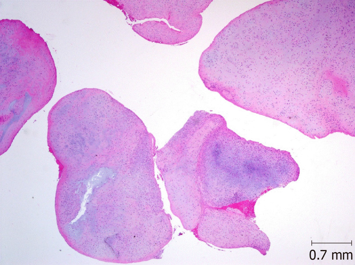
After osteochondral loose bodies had been fixed repaired tissues deteriorated significantly in accordance with duration of fragment isolation periods. We histologically examined 84 loose bodies and 9 related lesions synovial membrane nodules surgically removed from 24 joints of 24 patients with osteoarthrosis. Histologically based analyses of the nature and origin of loose bodies occurring in osteoarthrosis have been few and further study is warranted.
Histologically based analyses of the nature and origin of loose bodies occurring in osteoarthrosis have been few and further study is warranted. Osteochondromatosis SOC is a monoarticular synovial proliferative disease. This case highlights the importance of.
Three mechanisms for the generation. Attached bone could be seen from any of the three types of loose bodies but was rare in those specimens secondary to synovial osteochondromatosis. Learn vocabulary terms and more with flashcards games and other study tools.
Loose connective tissue histology slide with their identifying features 2. However in some cases even when. The loose bodies of the posterior ankle extra-articular space proper are removed to gain access to other parts of the posterior ankle.
Histologically based analyses of the nature and origin of loose bodies occurring in osteoarthrosis have been few and further study is warranted. It is a rare disease which presents as multiple cartilaginous nodules in synovial joints bursae or tendon. Intra-articular loose bodies can result from a variety of pathological processes.
Histologic classification of loose bodies in osteoarthrosis It is well known that loose bodies grow from proliferation of cartilage without blood supply in the joint cavity and that enchondral ossification is able to develop only under the condition of having a blood supply. Tissues are collections of cells that perform a similar function. Histologically based analyses of the nature and origin of loose bodies occurring in osteoarthrosis have been few and further study is warranted.
The imaging reports of the loose bodies suggested it to be synovial chondromatosis and histopathology studies confirmed this diagnosis. We histologically examined 84 loose bodies. Methods We histologically examined 84 loose bodies and 9 related lesions synovial membrane nodules surgically.
Additional histologic findings include papillary fibrocartilaginous metaplasia of the synovial surface sheets of coagulative necrosis without inflammation aggregates of hemosiderin-laden. Methods We histologically examined 84 loose bodies and 9 related lesions synovial membrane nodules surgically. Identification of dense regular connective tissue microscope slide with labeled diagram.

Qiao S Pathology Infarcted Appendix Epiploica Peritoneal Flickr

Histopathology Slide Of The Loose Body Showing Lobules Of Cartilage Download Scientific Diagram
Synovial Osteochondromatosis Of The Temporomandibular Joint A Case Report

A Rice Bodies Gross Specimen B Rice Bodies Microscopic Histology Download Scientific Diagram
Histological Examination Of The Loose Bodies Fibrous Connective Tissue Download Scientific Diagram
Webpathology Com A Collection Of Surgical Pathology Images

Autoamputated Adnexa Presents As A Peritoneal Loose Body Fertility And Sterility

Gross Finding Of Loose Bodies Several Loose Bodies Shown After Removal Download Scientific Diagram

Histological Features Of Oa The Normal Synovium Has A Thin 1e2 Cells Download Scientific Diagram

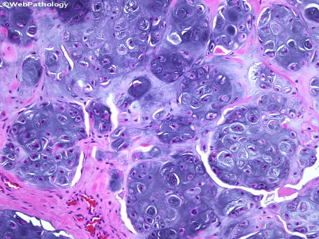
A Loose Bodies Showing Endochondral Ossification In The Hyaline Download Scientific Diagram
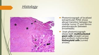
Pvns Synovial Chondromatosis Loose Bodies

Siu Som Histology Gi Histology Slides Loose Connective Tissue Medical Anatomy

Pdf Clinicopathological Study Of Gestational Trophoblastic Diseases Semantic Scholar

Qiao S Pathology Infarcted Appendix Epiploica Peritoneal Loose Body Or Peritoneal Mouse A Photo On Flickriver

Histologic Evaluation Of Osteochondral Loose Bodies And Repaired Tissues After Fixation Arthroscopy
Peritoneal Loose Body Flickr

Macro And Microscopic Findings Of The Loose Body A Extracted Loose Download Scientific Diagram

Figure 3 Arthroscopic Treatment Of A Case With Concomitant Subacromial And Subdeltoid Synovial Chondromatosis And Labrum Tear

Cartilage Proliferation Is Diagnosed During Histopathologic Evaluation Download Scientific Diagram


Comments
Post a Comment